Being told your dog has been diagnosed with an injury that involves surgery can be overwhelming, particularly if you don’t have all the facts.
As pet owners ourselves, we understand the thought of our beloved furry companions undergoing major surgery can be stressful.
We often find that once pet owners understand more about the disease process, what the surgery involves and what to expect with the recovery process helps them to make an informed decision and manage their expectations.
This blog is aimed at helping you, with our support, through this process.
What is cruciate ligament injury?
Cruciate ligament injury is very common in dogs and may involve a full or partial tear of the cranial cruciate ligament (called the anterior cruciate ligament or ACL in people). In dogs, rupture of the cranial cruciate ligament (CCL) is often associated with damage to other structures in the knee and arthritis.
How did my dog tear his ligament? He was normal yesterday!
In majority of the cases we see, owners tend to report that their dog was doing a normal activity such as chasing a ball in the park, or jumped off the couch, then suddenly appeared lame (not weight bearing on the hind leg).
Why is the knee so likely to be injured?
The knee joint is relatively unstable because there are no interlocking bones in the joint. Instead, the two main bones, the femur and tibia, are joined with several ligaments.
When severe twisting of the joint occurs, the most common injury is a rupture of the cranial cruciate ligament (CCL), which is one of two ligaments that cross over within the joint ensuring knee stability. When it is torn, instability occurs that allows the bones to move in an abnormal fashion in relation to each other.
How is it diagnosed?
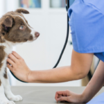
A full tear of the CCL can be diagnosed by detecting abnormal movement between the femur and tibia. This movement is called a drawer sign or cranial drawer. It can usually be demonstrated with the dog conscious, however, if there is severe pain, if the dog has very strong leg muscles, or is uncooperative, it may be necessary to give a general anaesthetic in order to examine the joint thoroughly.
Radiographs (x-rays) are always recommended to help diagnose concurrent disease, to formulate a more accurate prognosis and to establish the best treatment option.